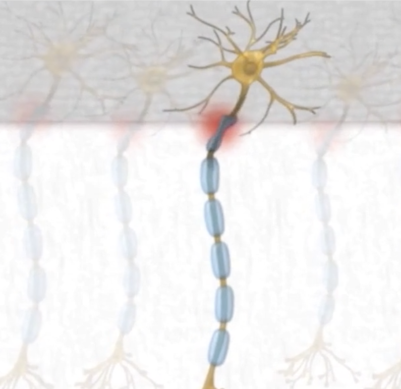
Brain injury – shear between white matter grey matter
When a brain injury occurs, especially in traumatic events like concussions or severe impacts, there can be a specific type of damage that affects the interface between white matter and grey matter. This damage is often referred to as shear injury or shearing.
Shear injuries involve the stretching, tearing, or disruption of nerve fibers (axons) at the boundary where white matter and grey matter meet. This boundary is called the gray-white matter junction. This area is particularly vulnerable to injury because of the differences in tissue density and structure between grey and white matter.

The forces exerted during a traumatic event can cause rapid acceleration or deceleration of the brain, leading to different parts of the brain moving at varying speeds. This discrepancy in movement can cause shearing forces that damage the delicate nerve fibers at this junction. As a result, axons may be stretched, twisted, or even torn, disrupting the communication pathways between different brain regions.
Shear injuries can lead to diffuse axonal injury (DAI), where widespread damage occurs to the axons throughout the brain. DAI can result in cognitive impairments, changes in consciousness, and various neurological deficits. This type of injury is commonly seen in severe traumatic brain injuries.
Understanding and addressing shear injuries in the brain are crucial for developing treatment strategies and rehabilitation plans for individuals who have experienced traumatic brain injuries. Techniques like neuroimaging (such as DTI – Diffusion Tensor Imaging) help in visualizing and assessing the extent of damage caused by shearing forces, aiding in diagnosis and treatment planning. Advancements have shown promise in detecting and mapping the integrity of white matter tracts, which can be affected by shear injuries. DTI measures the movement of water molecules in the brain and can indirectly assess the integrity of axonal fibers. But even with DTI, if it is available to you, it might not always be straightforward to attribute specific injuries to a precise event.
In the brain, white matter and grey matter are two types of tissue that play distinct roles in its structure and function.
- Grey Matter: Grey matter is primarily composed of neuronal cell bodies, dendrites, and synapses. It’s found in the outer layer of the brain (cerebral cortex) and in deeper brain structures. The grey color comes from the cell bodies and capillaries in this region. Grey matter is involved in processing and computation. Areas of the brain with high concentrations of grey matter are associated with various functions such as muscle control, sensory perception, memory, emotions, and decision-making.
- White Matter: White matter is composed mainly of axons, which are nerve fibers responsible for transmitting signals between different parts of the brain and spinal cord. These nerve fibers are surrounded by a fatty substance called myelin, which gives white matter its pale appearance. White matter forms the connecting pathways that facilitate communication between different regions of the brain. It enables the transmission of electrical signals between grey matter areas, allowing different parts of the brain to communicate and work together.
Dysfunction or damage to either white or grey matter can lead to various neurological conditions and cognitive impairments.
After a car accident, individuals may experience various types of dysfunctions, and visual, vestibular, and neck dysfunctions are common categories. These dysfunctions can often co-occur due to the complex nature of trauma. Here’s a breakdown of each:
- Visual Dysfunction:
- Symptoms may include blurred vision, double vision, saccades,light sensitivity (photophobia), difficulty focusing, and visual disturbances during movement.
- Visual problems can arise from direct head trauma, whiplash, or other injuries affecting the eyes or the brain’s visual processing centers.
- Accommodative disorders refer to difficulties in the eye’s ability to properly focus on objects at varying distances. This condition occurs when the eyes have trouble adjusting their focus from far to near or vice versa. It might result in blurred vision, eye strain, headaches, or difficulty concentrating on close-up tasks.
- Convergence insufficiency is a specific type of binocular vision disorder where the eyes have trouble working together when focusing on nearby objects. Individuals with this condition may experience double vision, eye strain, difficulty reading, and discomfort when doing tasks that require focusing on close objects for an extended period.
- Saccadic dysfunction refers to problems with the rapid, precise movements of the eyes when shifting gaze from one point to another- like looking at pictures in a film strip, verses watching a fluid video. Saccades are quick, jerky movements the eyes make to focus on different objects in the visual field. Dysfunction in these movements can lead to difficulties in tracking objects, reading, and following visual cues swiftly and accurately.
- Vestibular Dysfunction:
- Symptoms may include dizziness, vertigo (a spinning sensation), imbalance or unsteadiness, nausea, and sensitivity to motion or head movements. Noise sensitivity and Tinnitus
- Vestibular dysfunction often results from damage to the inner ear structures responsible for balance or disruption of signals between the inner ear and the brain.
- Neck Dysfunction:
- Commonly associated with whiplash, a neck dysfunction can result from the sudden back-and-forth motion of the head during a car accident.
- Symptoms may include neck pain, stiffness, reduced range of motion, headaches, and sometimes radiating pain to the shoulders or arms,
It’s important to note that these dysfunctions can interact and exacerbate each other. For example, neck pain and stiffness, Cervical instability, can contribute to visual symptoms – pain behind eyes, flashing lights in the brain, noise sensitivity, Tinnitus, nerve pain, chemical radiculopathy, heart issues, blood flow to the brain, balance issues and vice versa.
